Overview
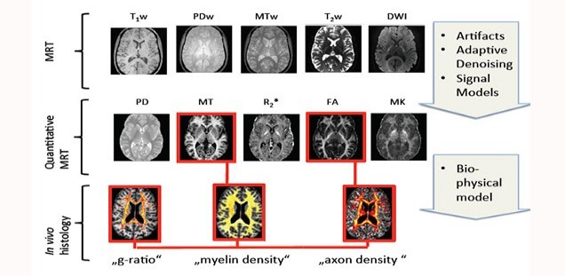
Understanding basic mechanisms underlying normal and diseased human brain developments crucially depends on reliable knowledge of its anatomical microstructure and connectivity. Our long-term goal is to estimate microstructural properties non-invasively in living subjects using biophysical models of the MRI signal.
We are developing novel methods for fusion of high-resolution quantitative MRI (qMRI) to enable MRI-based in vivo histology (hMRI) in the brain and spinal cord. The main focus is to develop valid MRI metrics that characterize key microstructure properties in the brain as known from ex vivo histology (e.g. cell size, myelin density, fiber density, and the g-ratio i.e. the ratio between inner and outer fiber diameter).
To achieve these goals, we are currently working on three projects:
Basic science/Clinical research: Non-invasive staining of tissue microstructure in temporal lobe epilepsy using in- vivo MRI (Funded by the ERC)
Completed projects:
Basic science: Validating MRI-based in vivo histology by comparison with ex vivo histology (Funded by the DFG Emmy Noether Program )
Clinical research: Understanding the microstructural mechanisms of spinal cord injury (Funded by EU ERA-NET Neuron )
Connectomics: In vivo and ex vivo characterization of the human microstructural connectome (Funded by DFG SPP 2041 ).
Marie Skłodowska-Curie Fellowship (MSCA-IF-2015): Mesoscopic characterization of human white-matter: a computational in-vivo MRI framework .
For a full publication list see: Siawoosh Mohammadi
- Staff
- Research topics
- Key Publications
- Funding
- Collaborations
- Vacancies
-
Staff
Principal Investigator
 Dr.Siawoosh MohammadiTelefonE-Mail
Our research group is based at two universities: the UKE and the University of Lübeck.The group members from Hamburg are:
Sophie Otte
The group members from Lübeck are:
Björn Fricke
Laurin Mordhorst
Jan Malte Oeschger
Nina Lüthi
Laura Bogs
Jan Meyer
Alumni
Behnam Ashtarayeh
Tobias Streubel
Gergely Dávid
Dr. Isabel Ellerbrock
Dr. Mohammad Hassan Farsbaf Shaker
Joshua Engels
Sara Holm
Hatice-Kübra Ögreten
Annkristin Lange
Christoph Nicolai
Dr. Thibault Tabarin
-
Research topics
- High-resolution quantitative MRI (qMRI) and in vivo histology (hMRI)
- Artifact correction and robust measurement of qMRI and hMRI metrics ( ACID toolbox )
- Neuroscience: structural and microstructural mechanisms of long-term nociception and pain
- Clinical research: robust biomarkers for spinal cord injury
-
Key Publications
High-resolution quantitative MRI and MRI-based in vivo histology
Mohammadi S, Carey D, Dick F, Diedrichsen J, Sereno MI, Reisert M, Callaghan MF and Weiskopf N (2015), Frontiers in Brain Imaging Methods, Whole-brain in-vivo measurements of the axonal g-ratio in a group of 37 healthy volunteers , 9: 00441, doi: 10.3389/fnins.2015.00441.
Mohammadi S, Tabelow K, Ruthotto L, Feiweier T, Polzehl J, Weiskopf N (2015) High-resolution diffusion kurtosis imaging at 3T enabled by advanced post-processing . Frontiers in Brain Imaging Methods 8:427,
N Weiskopf, S Mohammadi, A Lutti, MF Callaghan (2015) Advances in MRI-based computational neuroanatomy: from morphometry to in-vivo histology , Curr Opin Neurol. 28(4):313-22., doi: 10.1097/WCO.000000000000022
Edwards LJ, Pine KJ, Ellerbrock I, Weiskopf N, Mohammadi S (2017) NODDI-DTI: Estimating Neurite Orientation and Dispersion Parameters from a Diffusion Tensor in Healthy White Matter . Front Neurosci 11:720. doi: 10.3389/fnins.2017.00720.
Artifact correction and robust qMRI
Weiskopf N, Callaghan M, Josephs O, Lutti A, Mohammadi S (2014) Estimating the apparent transverse relaxation time (R2*) from images with different contrasts (ESTATICS) reduces motion artifacts . Brain Imaging Methods 8:278, doi: 10.3389/fnins.2014.00278.).
Mohammadi S, Möller HE, Kugel H, Müller DK, Deppe M (2010) Correcting eddy current and motion effects by affine whole-brain registrations: evaluation of three-dimensional distortions and comparison with slicewise correction. Magn Reson Med 64:1047–1056.
Structural changes associated with short-term sensitization in long-term nociceptive stimulation
Ellerbrock I, Mohammadi S (*), May A(*) (accepted) A model-based approach identifies strong and moderate sensitizers in a longitudinal pain paradigm In Proceedings of the 22nd Human Brain Mapping meeting, Geneva (2016); (*) shared senior authorship.
Robust spinal Cord Diffusion MRI
Mohammadi S, Freund P, Feiweier T, Curt A, Weiskopf N (2013a) The impact of post-processing on spinal cord diffusion tensor imaging . Neuroimage 70:377–385
David G, Freund P, Mohammadi S (accepted) The efficiency of retrospective artifact correction methods in improving the statistical power of between-group differences in spinal cord DTI . NeuroImage.
-
Funding
Secured Funding (last 5 years)
2017-2022 Emmy Noether Grant, German Research Foundation (DFG). Grant: MO 2397/4-1 .
2018-2020 Priority Programme Computational Connectomics (SPP 2041), German Research Foundation (DFG). Grant: MO 2397/5-1 ).
2017-2020 ERA-Net NEURON Cofund, European Unions Horizon 2020, Grant: No.88: hMRIofSCI
2015-2019 Co-PI on International Foundation for Research in Paraplegia grant (~185k) for a PhD position (Gergely David) for investigation of trauma induced structural spinal changes in collaboration with Patrick Freund in Zürich (Grant: P158 MRI).
2015-2017 Marie Curie Postdoctoral Fellowship, European Unions Horizon 2020, Grant: 658589 - MWMI.
2012-2014 DFG Postdoctoral Fellowship, Deutsche Forschungsgemeinschaft (DFG), Grant: MO 2397/1-1.
2013 UCL-ZNZ collaboration grant for laying the foundation of a larger multi-center grant between UCL and Zentrum für Neurowissenschaften Zürich (ZNZ).
-
Collaborations
MR Physics
Nikolaus Weiskopf
Department of Neurophysics, Max Planck Institute for Human Cognitive and Brain Sciences, Leipzig, GermanyMartina Callaghan
Wellcome Trust Centre for Neuroimaging, UCL Institute of Neurology,
University College London, UKMathematics and Computer Science
Lars Ruthotto
Department of Mathematics and Computer Science,
Emory University, Atlanta, USAKarsten Tabelow and Joerg Polzehl
Stochastic Algorithms and Nonparametric Statistics,
Weierstrass Institute for Applied Analysis and Stochastics, Berlin, GermanyPatrick Freund
Spinal Cord Injury Center Balgrist, University Hospital Zurich,
University of Zurich, SwitzerlandMarko Lindner
Institut für Mathematik,
Technische Universität HamburgJan Modersitzki
Institute of Mathematics and Image Computing,
University of LübeckHistology and Clinics
Patrick Freund
Spinal Cord Injury Center Balgrist, University Hospital Zurich,
University of Zurich, SwitzerlandMarkus Morawski
Paul Flechsig Institute for Brain Research,
University of LeipzigGabriele Rune and Lars Fester
Institute of Neuroanatomy,
Medical Center Hamburg-EppendorfKlaus Püschel
Department of Legal Medicine,
Medical Center Hamburg-Eppendorf
-
Vacancies
There are ongoing employment opportunities for doctoral students and post-docs. On a speculative application with motivation letter and CV to s.mohammadi@uke.de we look forward.